

From the time of conception, your baby goes through an intricate and complicated process of development. Your pregnancy’s first few weeks are critical, laying your baby’s foundation for a healthy and successful full-term birth. In the meantime, if you’ll soon be looking at the 5-week ultrasound pictures, what can you see?
An ultrasound scan at five weeks is taken to assess your baby’s development in the womb. The embryo is only the size of a pinhead or a sesame seed at this stage and resembles a tadpole, not a human baby. On the pictures, you’ll see a gestation sac within, which is one or two yolk sacs if you’re expecting multiples.
Due to the complexity of this initial stage of your pregnancy, it’s when difficulties are most likely to arise. Your doctor uses tests to assess the viability of your embryo, including 5 weeks ultrasound pictures that visualize your baby’s development. Let’s take a look at what you can expect to see in your baby’s first photo shoot, so to say.
What Happens to My Baby at Five Weeks of Pregnancy?
After five weeks of being pregnant, your body has undergone some rapid changes. Some of these you’re still coming to terms with. There won’t be much noticeable difference on the outside, but your uterus is already working overtime to nurture the growing embryo. One month and a week mark the phase where developments like heart, blood vessels, brain, and spinal cord take place.

At this stage, your baby’s nervous system is also developing, and that requires an abundance of micro and macronutrients. These include minerals, vitamins like B9, and iron. Other body features that start growing at five weeks are cardiovascular and respiratory systems and muscle tissue. All that growth is triggered by an influx of pregnancy hormones that cause your body and behavior affected.
Five weeks is the period you’ll most certainly be experiencing the worst of your pregnancy symptoms. You’ll suffer from headaches and nausea with constant mood or behavioral changes. Fatigue and lethargy are prevalent when doing anything at this stage.
The tests run at this level will confirm the presence of your baby, and whether the embryo is growing at an expected rate. Your doctor will look for abnormalities within your uterus, observing the amniotic, gestation, and yolk sacs as indicators of a healthy pregnancy. That information is also used to estimate your pregnancy’s age and to give a reliable expected date of delivery.
What Will I See on My 5-Week Ultrasound Pictures?
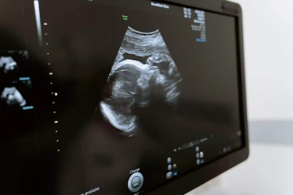
During the first trimester, which lasts from conception to the end of three months a, uterine gestation sac has formed. Inside this space is a yolk sac, embryonic disk, and amnion. However, these structures are too tiny to be observed using sonography. At this stage, your sonographer may point out a tiny white and curled object, which is the embryo.
Surrounding the embryo, you’ll see a yolk sac that nourishes it. That small white circle is where your baby’s first blood cells are manufactured. The gestation sac is the large black area on your 5week ultrasound pictures, which contains amniotic fluid. Its measurements are initially 0.2 to 0.3 inches, but it grows at a rate of 0.113 inches in diameter daily.
There may also be flickers of a heartbeat, but it’s common to miss one at five or even six weeks of pregnancy. Sometimes, you’re not on the actual gestational age’s fifth week, or your human chorionic gonadotropin or hCG levels are insufficient.
It’s the same hormone that your home pregnancy kit detected in your urine when you tested positive. At five weeks of pregnancy, the level should exceed 2,500 or 3,500 mIU per ml of serum if you’re to see anything on the ultrasound pictures.
Which Ultrasound Diagnostic Technique Offers the Best Results at Five Weeks of Pregnancy
During early pregnancy, you’ll normally have an ultrasound scan around the 12th week. Early scans, some as soon as four or five weeks can be requested by your doctor are you’re bleeding or experiencing pain. These tests also act to give a reliable expected day of delivery, especially if you’re unsure of your last monthly period or LMP dates.
At this stage, your obstetrician-gynecologist will recommend either the trans-abdominal or TAU and transvaginal ultrasound or TVU diagnostic tools. While the former is non-evasive, it offers an abdominal and pelvis panorama. The latter requires the insertion of a vaginal probe, providing a closer inspection of your uterus.
At around five weeks gestation, the TAU can’t provide reliable pregnancy diagnosis, while the TVU detects them earlier. The ultrasound device is also used to check the placement of the embryo and that of the placenta. Thats especially if you’re expecting twins.
An ultrasound scan in this period also helps to provide markers of chromosomal abnormalities within your uterus. These can include increased fetal nuchal translucency or absence of the fetal nasal bone which can indicate Down syndrome.
Why Can’t I See My Baby on Five-Week Ultrasound Pictures?
Your sonographer can explain what you’re looking at as it may be unclear what you’re seeing on your 5-week ultrasound pictures. There won’t be much to see in terms of facial or bodily features. The presence of a yolk sac points to an intrauterine pregnancy as opposed to an ectopic one.
By five weeks and four days, the yolk sac is visible, or more than one in the case of multiples. Maternal or identical twins have yolks that share a gestation sac; there’ll be two sacs if yours are fraternal. You’ll also see spaces and cavities, also called lacunary structures at the site of embryo implantation. Next to these sacs is the embryonic pole from which cardiac activity can sometimes be seen.
Another reason to not see a gestation sac in your five-week ultrasound pictures is miscarriage which is common at this stage due to failing hCG levels. It’s what happens when the zygote fails to form a viable embryo and can be indicated by vaginal bleeding. It’s not a unique indicator, and spotting could be pointing to something else altogether.
Conclusion
You can expect to see something in your five-week ultrasound pictures. But it’s not anything to post on social media or hang on your wall. If there’s a reason for your doctor to suspect abnormalities, more tests will be conducted as your pregnancy progresses and early treatment is sought. Keep up with your recommended prenatal appointments and vitamin intake. You can wait to see more of your unborn baby’s features in the 12-week scan.

I’m Cathrine and I’m a 39-year-old mother of 3 from Utica, New York. And I’m extremely happy you’ve come to visit my hide-out on the web. Here I post about everything related to family-life and usually it will involve babies and lessons I’ve learned over the years from experts, friends, and my own mistakes. So hopefully you will find what i write fun and informational!
